Home
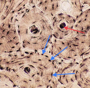
/ Cross Section Of A Compact Bone : Structure and Function of Joints | Musculoskeletal Key - (b) in this micrograph of the osteon, you can clearly see the concentric lamellae and central canals.
Cross Section Of A Compact Bone : Structure and Function of Joints | Musculoskeletal Key - (b) in this micrograph of the osteon, you can clearly see the concentric lamellae and central canals.
Cross Section Of A Compact Bone : Structure and Function of Joints | Musculoskeletal Key - (b) in this micrograph of the osteon, you can clearly see the concentric lamellae and central canals.. In the center of each osteon is the central canal, a space that houses blood vessels and nerves that supply bone. There are two ways to study bone histology. The outlined area is a cross section of an osteon of compact bone. In three dimensions an osteon is cylindrical in shape. This model shows a cross section of compact bone.
Compact bones make up 80 percent of the human skeleton; As the names suggest compact bone looks compact and the spongy bone looks like sponges. Compact bone, also known as cortical bone, is a denser material used to create much of the hard structure of the skeleton. An estimated 10 percent of an adult's skeleton is replaced each year. A cross section of a compact bone shows concentric circles called lamellae.
Muscular and Skeletal Systems - Histology from uta.pressbooks.pub As the names suggest compact bone looks compact and the spongy bone looks like sponges. Remodeling allows the body to fix damaged sections, reshape the skeleton during growth, and regulate calcium levels. This model shows a cross section of compact bone. A central tube called a haversian canal typically runs in the same path as the length of the bone. These are abundant and characteristic of compact bone. (b) in this micrograph of the osteon, you can clearly see the concentric lamellae and central canals. Cross section of the compact bone. Bone must be decalcified (by exposure to strong acids) so it can be cut into thin sections.
The remaining material is mostly collagen.
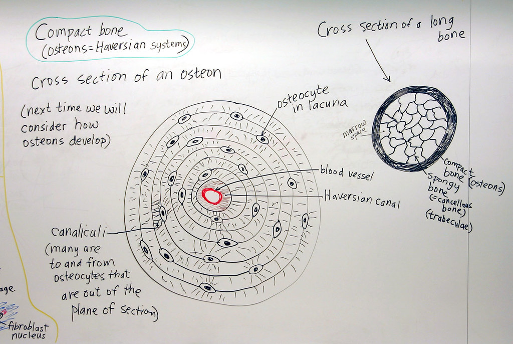
Structures and bone areas in column b, and use them to color the coding. These are abundant and characteristic of compact bone. Osteocyte processes lie in tiny canals (canaliculi) in the bone matrix. The remaining material is mostly collagen. Cross section of compact bone. Concentric layers of bone cells (osteocytes). Observe that the matrix of the bone is deposited in concentric layers that are called lamellae (5). The outlined area is a cross section of an osteon of compact bone. They build the entire picture, improve your understanding, consolidate the information and facilitate recall. Most bones contain both compact and spongy bone. A central tube called a haversian canal typically runs in the same path as the length of the bone. Cross section of the compact bone. Select different colors for the.
The outlined area is a cross section of an osteon of compact bone. Compact bone (cross section of dried bone). Compact bone is very different from the other tissues you have seen. (b) in this micrograph of the osteon, you can clearly see the concentric lamellae and central canals. Cross section of the compact bone.
Doctors Gates: Compact and Spongy Bones from 1.bp.blogspot.com (micrograph provided by the regents of university of michigan. Spongy bone and compact bone. The two layers of compact bone and the interior spongy bone work together to protect the internal organs. An estimated 10 percent of an adult's skeleton is replaced each year. The remaining material is mostly collagen. These are mostly compacted bone with little marrow and include most of the bones in the limbs. I am sure they have a higher strength vs weight ratio. Compact bone (cross section of dried bone).
The spongy and compact bone tissue in the cross section of a skull bone.
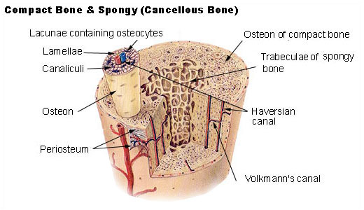
Canaliculi allow the passage of interstitial fluid between the central canal and the lacunae housing osteocytes. Magnification view of compact bone tissue. Most but not all human bones have circular cross sectional shapes. In a cross section of a bone we can see two types of bone tissue: The innermost layer of membrane is made up of. Also called cortical bone, the compact variety usually features a haversian system, or cylindrical unit within the structure. Remodeling allows the body to fix damaged sections, reshape the skeleton during growth, and regulate calcium levels. Dry bone is cut and polished before mounting on a slide. Compact bone (cross section of dried bone). Spongy bone is the osseous tissue, which fills the interior cavity of bones, consisting of mineralized bars called trabeculae. The connection point for the periosteum. These are mostly compacted bone with little marrow and include most of the bones in the limbs. A cross section of a human long bone.
The remainder is spongelike cancellous bone. Dry bone is cut and polished before mounting on a slide. The basic unit of structure in this type of bone is the haversian system, or osteon. Compact bones make up 80 percent of the human skeleton; This is a short tutorial using blender 2.8 that shows how to create a bone cross section and using images to create the textures.
Cartilage and Bone: Compact Bone | A hand drawn sketch by ... from c2.staticflickr.com In three dimensions an osteon is cylindrical in shape. This is a cross section through decalcified bone. The outlined area is a cross section of an osteon of compact bone. A cross section of a compact bone shows concentric circles called lamellae. In the center of each osteon is the central canal, a space that houses blood vessels and nerves that supply bone. Bone must be decalcified (by exposure to strong acids) so it can be cut into thin sections. This is a short tutorial using blender 2.8 that shows how to create a bone cross section and using images to create the textures. Osteocyte processes lie in tiny canals (canaliculi) in the bone matrix.
Osteocyte processes lie in tiny canals (canaliculi) in the bone matrix.
They build the entire picture, improve your understanding, consolidate the information and facilitate recall. As the names suggest compact bone looks compact and the spongy bone looks like sponges. Their course follows the main axis of long bone. Spongy bone is the osseous tissue, which fills the interior cavity of bones, consisting of mineralized bars called trabeculae. Remodeling allows the body to fix damaged sections, reshape the skeleton during growth, and regulate calcium levels. A cross section of a compact bone shows concentric circles called lamellae. I am sure they have a higher strength vs weight ratio. Also called cortical bone, the compact variety usually features a haversian system, or cylindrical unit within the structure. In the center of each osteon is the central canal, a space that houses blood vessels and nerves that supply bone. The spongy and compact bone tissue in the cross section of a skull bone. These are abundant and characteristic of compact bone. Magnification view of compact bone tissue. The innermost layer of membrane is made up of.
 in this micrograph of the osteon, you can clearly see the concentric lamellae and central canals.)